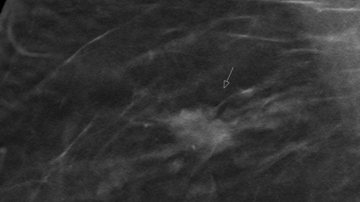
A large German study has found that digital breast tomosynthesis detects breast cancer more effectively than standard digital mammography during routine screening. Researchers from the University of Münster and the University Hospital Münster conducted the TOSYMA trial, analyzing nearly 100,000 women aged 50 to 70 across 17 screening centers in North Rhine-Westphalia and Lower Saxony between 2018 and 2020.
Study Design and Methods
Participants were randomly assigned to either tomosynthesis screening or digital mammography. In the tomosynthesis group, multiple low-dose X-ray images were taken from different angles, allowing reconstruction of cross-sectional breast images up to one millimeter thick. This technique reduces the likelihood of tumors being obscured by overlying tissue. The other group received standard digital mammography.
The study was embedded within Germany’s nationwide, quality-assured mammography screening program, ensuring it took place under real-world conditions rather than a controlled laboratory setting. To track interval cancers—those diagnosed between regular screenings—researchers cross-referenced participant data with cancer registries covering over 95% of new breast cancer cases in the region. The suspected cases were reviewed through a multistep process involving staff at the cancer registries and reference centers, as well as an independent data safety committee, all of whom remained unaware of each participant’s assigned group throughout the evaluation.
The first analysis of the TOSYMA-1 study, published in 2022, showed that tomosynthesis detects a significantly higher number of invasive breast cancers—specifically 7.1 versus 4.8 per 1,000 women screened. However, it remained unclear whether tomosynthesis was also clinically more effective. The second analysis (TOSYMA-2) addresses this issue by comparing interval cancers in both groups. In the tomosynthesis group, 4.5 out of 10,000 women were diagnosed with invasive cancer during the first year after receiving a normal screening result. In the mammography group, this rate was 6.3.
Key Findings on Interval Cancers
Interval cancers, which are typically more aggressive and have a poorer prognosis than those found during scheduled screenings, serve as a key measure for early detection effectiveness. In the first year after a normal screening result, the invasive breast cancer diagnosis rate was 29% lower in the tomosynthesis group compared to the mammography group.
Over a two-year follow-up period, the rate of invasive interval cancers was 14.2 per 10,000 women in the tomosynthesis group versus 18.6 per 10,000 in the mammography group, representing a 24% reduction. However, this difference was not statistically significant, which researchers partially attributed to the COVID-19 pandemic disrupting recruitment and resulting in approximately 20,000 fewer participants than originally planned.
The benefits were most pronounced among women returning for a second screening round. Their two-year interval cancer rate was 13.5 per 10,000 in the tomosynthesis group, while first-time participants showed nearly identical rates between groups (18.8 per 10,000).
Additional Clinical Outcomes
Beyond detecting fewer interval cancers, the tomosynthesis group also demonstrated advantages in tumor characteristics. Interval cancers diagnosed in this group were less likely to be locally advanced or to have spread to axillary lymph nodes, both indicators of more advanced disease at diagnosis.
Program sensitivity, defined as the proportion of invasive cancers detected during the screening examination itself, was higher in the tomosynthesis group at 83.5% compared to 72.3% in the mammography group. Specificity, the ability to correctly identify women without cancer, remained similar across both groups.
“Our study decisively strengthens the scientific basis for tomosynthesis in breast cancer screening,” said Professor Stefanie Weigel from the Department of Radiology at the University of Münster and UKM. “It expands and enhances the previously limited body of research on follow-up after tomosynthesis screening.”
The research team plans to analyze cancers detected during subsequent routine follow-up screenings to determine whether tomosynthesis reduces advanced tumor stages more effectively than mammography over time. Professor Walter Heindel, the lead investigator, emphasized that this next phase will provide further insight into the long-term benefits of the technology. Using digital breast tomosynthesis in the breast cancer screening program could prevent around a quarter of invasive interval cancers that occur within two years after a digital mammogram.
The findings were published in The Lancet Oncology, marking the largest comparative study of the two diagnostic methods conducted worldwide to date. With approximately 75,000 new breast cancer cases diagnosed annually in Germany, making it the most common cancer among women, improvements in early detection carry significant public health implications. The study’s results highlight the potential of tomosynthesis to enhance screening accuracy and patient outcomes in a real-world setting.
The research was published as Stefanie Weigel et al., Invasive interval breast cancer rates after digital breast tomosynthesis plus synthesised mammography versus digital mammography screening (TOSYMA): 24-month follow-up of the multicentre, open-label, randomised, controlled, superiority trial, The Lancet Oncology (2026). DOI: 10.1016/s1470-2045(26)00341-4.